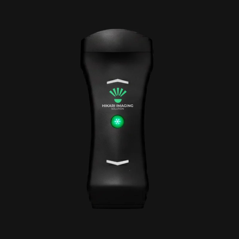
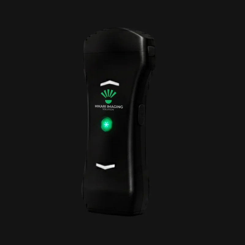
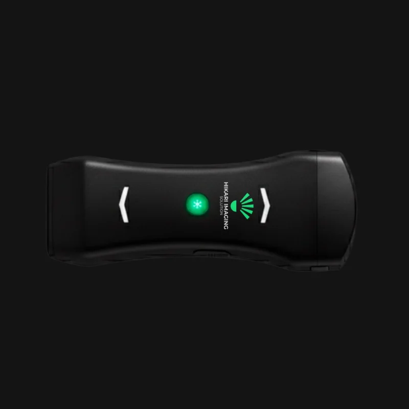
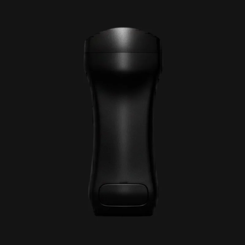
The HK-3 from Hikari Imaging Solution is a compact wireless ultrasound probe weighing just 220 g. Measuring 160 mm x 70 mm x 30 mm, it is designed for maximum portability and ease of use across a variety of clinical settings. The probe features a 128-element design and supports wireless charging.
It offers a wide range of operating modes, including B-mode, Color Flow (CF) mode, M-mode and Pulsed-Wave (PW) Doppler. With applications in abdominal, cardiac, vascular, obstetric and gynecological imaging, the HK-3 stands out as a versatile diagnostic tool.
It is compatible with Windows and Android devices (including PCs, laptops, tablets and smartphones), enabling flexible use and real-time imaging. It also includes a complete package of specialized measurements and calculations for a range of medical fields.
Designed for mobility and precision, the HK-3 combines high-end imaging with true portability.
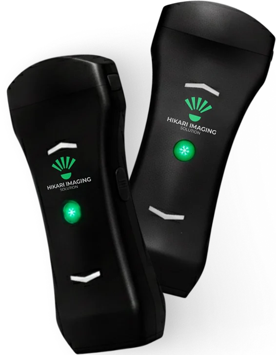

THI/iTHI, Noise (Speckle) Reduction and Spatial Compound Imaging.

Works with Windows PCs, notebooks, tablets and Android devices.

Built-in rechargeable battery with wireless charging.

DICOM compatible; export to BMP, JPG, PNG, TIFF, WMV and MP4.

B, M, Doppler (PW, CF, PDI, DPDI, TDI), Wide View, Needle Enhancement.

160 × 70 × 30 mm and just 220 g – designed for mobility.
From routine exams to specialized measurements, the Hikari HK-3 adapts seamlessly to diverse clinical needs.

Tissue Doppler imaging, LA/Ao ratio, ejection fraction, cardiac output and valve assessments.

IMT, % stenosis, vessel diameters, flow analysis and PI/RI indices.

Liver, gallbladder, pancreas, kidneys, spleen and vascular structures.

Hip dysplasia analysis (Graf method) and femoral head coverage.

Fetal biometry (BPD, HC, AC, FL, CRL, NT, AFI, EFW), multiple-pregnancy follow-up and uterine/ovarian volumes.

Prostate, bladder, residual urine, thyroid, testicles and PSAD calculations.
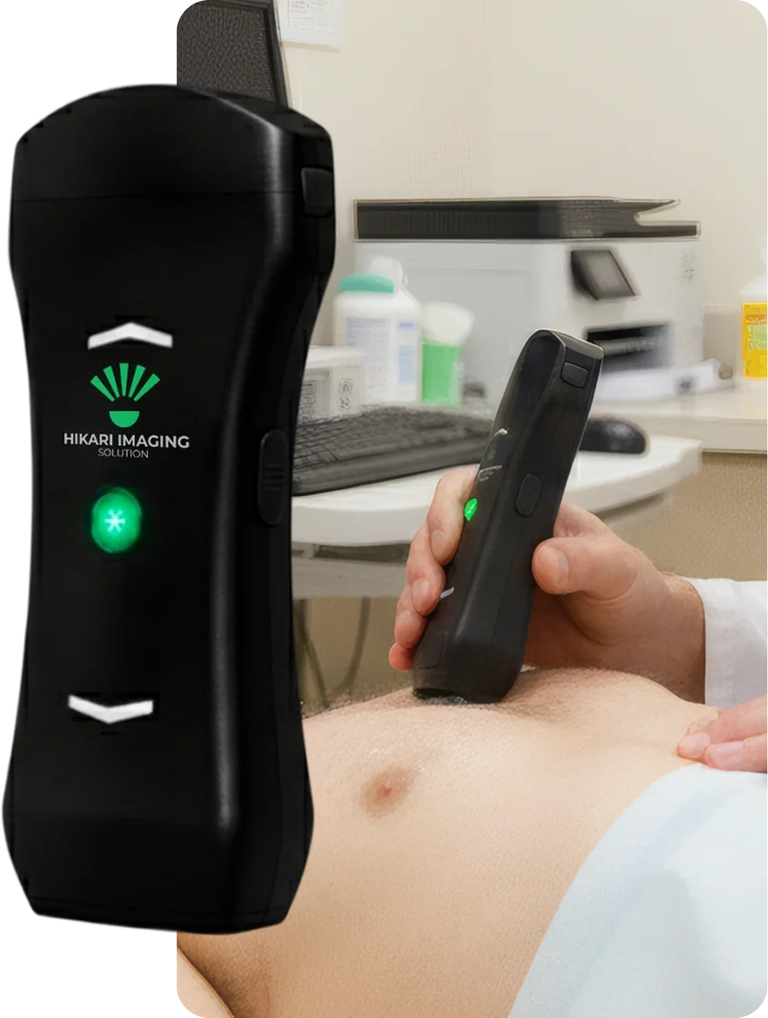


Request more information or a personalized demo. Our technical team is ready to help.
Book a demo
©2026 Hikari Medical Solutions. All rights reserved.